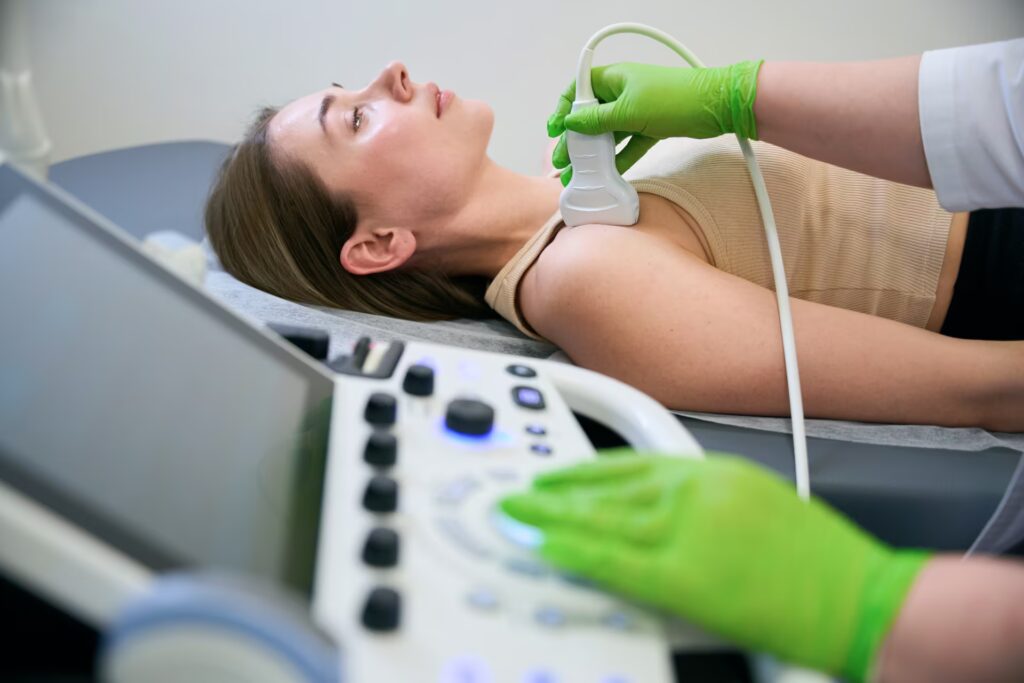
Ultrasound Scans
An ultrasound scan is a non-invasive imaging technique that uses high-frequency sound waves to visualise internal structures, such as muscles, tendons, joints, or organs. In physiotherapy, it’s often used during an initial assessment to evaluate musculoskeletal injuries, like sprains or muscle tears, providing real-time insights into tissue condition. This helps physiotherapists design targeted treatment plans, monitor healing, and assess inflammation or abnormalities without radiation exposure. Ultrasound aids in accurate diagnosis, guiding interventions like exercise or manual therapy.
For someone undergoing their first ultrasound scan, the experience is straightforward and painless. You’ll lie on an examination table, and the physiotherapist or technician will apply a cool, water-based gel to the skin over the area being examined. This gel enhances sound wave transmission. A handheld device, called a transducer, is gently moved across the skin, emitting sound waves that reflect off tissues and create detailed images on a screen. The procedure typically lasts 10-20 minutes, with no discomfort beyond mild pressure from the transducer. You may hear a faint pulsing sound from the machine. Afterward, the gel is wiped off, and you can resume normal activities immediately. Results are often discussed during or after the assessment to inform your treatment plan.

